Eye Anatomy
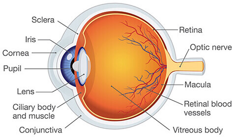
In order to fully grasp the effects some eye diseases have on your vision, it’s helpful to have a basic understanding of eye anatomy. The primary parts of the eye are described in more detail, below.
Cornea
The cornea is the clear, dome-shaped front part of the eye. Its purpose is to focus light as it enters the eye, so you can see. Corneal tissue is comprised of five layers — the epithelium, Bowman’s membrane, the stroma, Descemet’s membrane, and the endothelium. The cornea receives nourishment from tears, which are produced by the lacrimal glands. Common conditions that affect the cornea are:
- Allergies – Allergy symptoms often include redness, itching, tearing, burning, or stinging of the cornea. Over-the-counter antihistamine medication or eyedrops can help relieve these symptoms.
- Keratitis – Noninfectious keratitis is an inflammation of the cornea, usually caused by a minor injury or from wearing contact lenses for an extended period of time. Infectious keratitis is caused by bacteria, viruses, fungi, parasites, or contact lenses. Minor corneal infections are typically treated with antibacterial eye drops or steroid drops to reduce inflammation.
- Dry Eye – Dry eye syndrome is usually characterized by a stinging, burning, or scratchy sensation. The condition develops when the eye produces fewer tears or lower quality tears, which results in a lack of lubrication for the cornea. Treatment for mild cases of dry eye includes artificial tears, prescription eye drops, or warm compresses. More severe cases of dry eye may benefit from advanced treatment such as punctal plugs, BlephEx, or tear duct surgery.
- Keratoconus – Keratoconus is a gradual thinning of the cornea, which causes the middle of the eye bulge outward. The abnormal curvature of the cornea can cause blurry vision, nearsightedness, or astigmatism. In its early stages, keratoconus is treated with prescription eyeglasses or contacts. Severe cases of keratoconus can be treated with procedures such as corneal crosslinking or a corneal transplant.
- Conjunctivitis – Conjunctivitis is a minor infection of the conjunctiva, the thin, clear layer of tissue that lines the inner surface of the eyelid. While commonly caused by a viral or bacterial infection, conjunctivitis can also develop as a result of an allergic reaction to pollen, smoke, chlorine, cosmetics, or contact lenses. Treatment typically includes antibiotic eye drops, cool compresses, and/or artificial tears. Conjunctivitis is especially common in children.
- Pterygium – Also known as “surfer’s eye,” a pterygium is an elevated triangular growth of tissue on the cornea. Pterygium is caused by prolonged exposure to wind, dust, and UV sunlight. Treatment depends on the size of the growth and its symptoms. Mild cases can be treated with steroid eye drops, while more severe forms of pterygia may require surgery.
Lens
The lens is a transparent, convex structure that sits behind the iris, the colored part of your eye. The lens is responsible for focusing the light rays that enter the eye through the cornea, so you can see at multiple distances. Common diseases that affect the lens include:
- Cataracts – Most cataracts are linked to aging. The common eye condition occurs when the lens of the eye becomes clouded with proteins, which results in blurry vision over time. The most effective way to treat cataracts is to remove the lens of the eye during cataract surgery and replace it with an intraocular lens (IOL).
- Presbyopia – An age-related form of farsightedness, presbyopia occurs when the lens of the eye loses its elasticity, which results in poor up-close vision. Middle-aged adults with presbyopia usually have difficulty reading fine print or seeing text messages. The common treatment for presbyopia includes progressive eyeglasses or contact lenses that provide clear vision at all distances. Refractive lens exchange (RLE) is a surgical procedure that’s essentially the same as cataract surgery, except there is no cataract present. During surgery, the lens is removed and replaced with a multifocal IOL or an accommodating IOL to restore near vision in patients with presbyopia.
Retina
The retina is the thin, light-sensitive layer of tissue that lines the back of the eye. The purpose of the retina is to receive light from the lens of the eye and convert it to neural signals that are sent to the brain. Damage to the retina can cause severe vision loss or permanent blindness. Common retinal diseases include:
- Diabetic Eye Disease – People with diabetes are more susceptible to retinal damage, in addition to glaucoma and cataracts. Diabetic retinopathy is a condition that causes swelling in the macula, an area of the retina responsible for central vision. Diabetic macular edema (DME) is a consequence of diabetic retinopathy.
- Retinal Detachment – As we age, the gel-like substance inside the eye, called the vitreous, changes in consistency. The fluid can collect underneath the retina and cause a hole or tear. If the vitreous liquid passes through the tear into the space behind the retina, the retina can become detached, causing the sudden appearance of floaters, flashes of light, reduced peripheral vision, blurry vision, or a curtain-like shadow in your visual field. Retinal detachment is considered a medical emergency — if left untreated, you can permanently lose your vision.
Optic Nerve
The optic nerve is a bundle of nerve fibers that transmit visual information from the retina to the brain. Located at the back of the eye, the optic nerve is an essential part of eye anatomy, as it is partly responsible for our ability to see. The most common eye condition that affects the optic nerve is glaucoma.
- Glaucoma – a group of diseases that affect the optic nerve, glaucoma develops when the level of pressure inside the eye increases. The high pressure causes the optic nerve to compress, which results in cell damage. While glaucoma progresses slowly over time, if left untreated, it can cause blurry vision or permanent blindness.
Related Pages