If your eye doctor suspects that you have “narrow” or “closed” angles, this means that the drainage channel of your eye is blocked or nearly blocked, placing you at high risk for elevated intraocular pressure and vision loss. This is called “narrow-angle” or “angle-closure” glaucoma.
An acute attack of angle-closure glaucoma is marked by very high eye pressure and complete blockage of the drainage channel in the eye. Symptoms include pain, red eye, and decreased vision.
To treat angle-closure glaucoma, your ophthalmologist will perform a laser peripheral iridotomy (LPI), creating a surgical opening within the upper part of the iris (the colored part of the eye) using a laser. This opening is typically so small that it cannot be seen with the naked eye. The opening in the iris allows fluid to flow from behind the iris through the opening, allowing the iris to fall back into a more normal position and opening the drain.
This laser treatment is always performed on an outpatient basis, often in the ophthalmologist’s office. The treatment will not improve your vision, but it can help prevent vision loss from a dangerous type of glaucoma. The side effects of the treatment can include the appearance of a “light streak,” a temporary rise in intraocular pressure, and inflammation.
Selective laser trabeculoplasty (SLT) is a laser surgical procedure used to help lower intraocular pressure (IOP) of patients with open-angle glaucoma. SLT is used to treat the eye’s drainage system, known as the trabecular meshwork -the mesh-like drainage canals that surround the iris. Treating this area of the eye’s natural drainage system improves the flow of fluid out of the eye, helping to lower the pressure.
The laser used in SLT works at very low levels. It treats specific cells selectively, leaving untreated portions of the trabecular meshwork intact. For this reason, SLT, unlike other types of laser surgery, may be safely repeated many times.
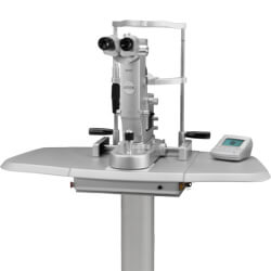
SLT is performed in the ophthalmologist’s office or an outpatient surgery center. The procedure usually takes about five to ten minutes. First, anesthetic drops are placed in your eye. The laser machine looks similar to the examination microscope that your ophthalmologist uses to look at your eyes at each office visit.
You will experience a flicker of light with each laser application. Most people are comfortable and do not experience any significant pain during the surgery, although some may feel a little pressure in the eye during the procedure.
Most people will need to have their pressure checked after the laser treatment since there is a risk of increasing IOP after the procedure. If this does occur, you may require medications to lower the pressure, which will be administered in the office. Rarely, the pressure in the eye increases to a high level and does not come down. If this happens, you may require surgery in the operating room to lower the pressure.
Most people notice some blurring of their vision after the laser treatment. This typically clears within a few hours. The chance of your vision becoming permanently affected by this laser procedure is very small.
Most patients can resume normal daily activities the day after laser surgery. You may need to use eye drops after the procedure to help the eye heal properly.
It will take several weeks to determine how much SLT will lower your eye pressure. You may require additional laser or glaucoma drainage surgery to lower the pressure if it is not sufficiently lower after the first laser treatment.
Most patients must continue to take the medication in order to control and maintain their IOP; however, surgery can lessen the amount of medication needed.
While some people may experience side effects from medications or surgery, the risks associated with these side effects should be balanced against the greater risk of leaving glaucoma untreated and losing your vision.
If you have glaucoma, and medications and laser surgeries do not lower your eye pressure adequately, your eye doctor may recommend a procedure called a trabeculectomy.
In this procedure, a tiny drainage hole is made in the sclera (the white part of the eye). The new drainage hole allows fluid to flow out of the eye into a filtering area called a bleb. The bleb is mostly hidden under the eyelid. When successful, the procedure will lower your intraocular pressure (IOP), minimizing the risk of vision loss from glaucoma. The surgery is performed in an operating room on an outpatient basis.
Some of the risks and complications from trabeculectomy surgery include the following:
Certain medications, called antimetabolites, were originally developed to help treat some kinds of cancer. These same medications have also been found to be helpful when used with some types of glaucoma surgery.
These medicines may be applied to the eye during or after the surgery to reduce the growth of scar tissue, a common cause of failure in glaucoma surgery. Mitomycin-C and 5-fluorouracil (5-FU) are the most commonly used antimetabolites for glaucoma surgery. When these antimetabolites are used with other medications that reduce inflammation, the success rate of surgery is greatly improved, especially for patients who are at high risk for excessive scarring.
While some people may experience side effects from medications or surgery, the risks associated with these side effects should be balanced against the greater risk of leaving glaucoma untreated and losing your vision.
Surgery may be necessary to treat glaucoma, but often it is not the best option. Less invasive treatments can be equally effective and are less intense. There is a group of procedures called minimally invasive glaucoma surgeries or MIGS. They provide more straightforward glaucoma treatments than traditional glaucoma surgery.
There are numerous MIGS procedures available to reduce your IOP and your use of medications. At The Medical Eye Center, Dr. Amy Hennessy performs all the MIGS procedures. She may use one of the following techniques to reduce your IOP:
All these procedures are effective, and each is beneficial for different situations. Dr. Hennessy will help you find the procedure best suited for your eyes.
iStent micro-bypass inserts two tiny devices into your eyes to reduce IOP. It can occur during cataract surgery or as a stand-alone procedure. The devices get implanted in your eye’s drainage network. They create two new pathways for eye fluid to flow through. These new pathways help keep IOP low and may reduce the number of glaucoma medications you need to take.
Goniotomy is a MIGS procedure that removes a small section of the trabecular meshwork to create a new outflow. It gives eye fluid a path to enter the collector channels of your drainage network. By creating a new outflow channel, goniotomy can help to reduce IOP. This reduction in IOP can limit your use of glaucoma medication and help protect your optic nerve.
Ab Interno Canaloplasty is a MIGS procedure similar to a traditional trabeculectomy. But, it is safer than conventional surgeries, with fewer risks and side effects. That does not mean it is less effective, though. Often, Ab interno canaloplasty is as effective as other more invasive glaucoma procedures.
It addresses all potential blocks in your eye’s outflow channels without damaging any tissue. Also, Ab interno canaloplasty does not use any stents or equipment. That means it reduces your IOP without leaving any foreign objects in your eye. Yet, it still lowers your need for glaucoma medication to keep your IOP at healthy levels.
The Hydrus microstent is an implantable device that gets placed in your eye during cataract surgery. It goes in your Schlemm’s Canal, which is the main drainage pathway out of your eye. Before your surgeon places your IOL, they put the microstent in using the incision in your cornea. The device then expands to open the canal, allowing eye fluid to drain naturally.
Trabectome removes tissue from your drainage network to open an outflow pathway. It uses an electrosurgical pulse to cauterize tissue in your Schlemm’s canal. Then your surgeon removes the cauterized tissue. It creates more space for fluid to enter the back of your eye and flow out towards your nasal passage. It also makes future glaucoma procedures more straightforward if they are necessary.
MIGS procedures provide gentle methods to reduce IOP and dependence on glaucoma medications. Dr. Hennessy will help you determine if you qualify for a MIGS procedure and which is best for you.
Schedule an appointment at one of our four locations. Determine if you qualify for a MIGS procedure to treat your glaucoma!