
Diplopia (double vision) caused by a problem with the muscles that control the movement of the eye or the nerves that stimulate those muscles.
The onset of double vision in adulthood should be brought to the attention of your ophthalmologist immediately to exclude the possibility of a tumor, aneurysm, or neurological problem. Two of the primary neurological conditions that could cause diplopia are microvascular cranial nerve palsy and myasthenia gravis .
Microvascular cranial nerve palsy is one of the most common causes of double vision in older people. It occurs more often in patients with diabetes and high blood pressure, when blood flow is blocked to one or more of the six eye muscles that control eye movement. Although there is no known treatment for microvascular cranial nerve palsy, the double vision may alleviated by placing a prism on your spectacles or wearing an eye patch.
Myasthenia gravis is a disorder characterized by muscle weakness, caused by a communication breakdown between the nerves and the muscles due to an autoimmune condition. It is most common in the muscles of the face, eyes, arms, and legs, and in those involved in chewing, swallowing, and talking. Double vision is one of the common indicators of myasthenia gravis. Though there is no known cure for myasthenia gravis, there are a number of treatment options to manage the condition, including medications, physical therapy and surgery. Early detection and treatment of myasthenia gravis is crucial to managing the condition and preventing serious problems with breathing or swallowing, which can require emergency care.
Microvascular cranial nerve palsy is one of the most common causes of double vision in older people. It occurs more often in patients with diabetes and high blood pressure and is often referred to as a “diabetic” palsy.
Microvascular cranial nerve palsy occurs when the blood flow is blocked to one or more of the nerves that control the eye muscles. Injury to the abducens nerve will cause your eye to not be able to move toward the outside. This creates double vision with side-by-side images. If the trochlear nerve is affected the double vision will be vertical (one image on top of another). And if the oculomotor nerve is affected there may be drooping of the eyelid along with double vision.
Although it is not clear what blocks the blood flow, diabetes and high blood pressure are often associated with this condition.
Symptoms of microvascular cranial nerve palsy include blurred or double vision, drooping of an eyelid, or enlarged pupil.
Although there is no known treatment for microvascular cranial nerve palsy, double vision may be treated by patching either eye or placing a prism in the patient’s spectacles. If the double vision persists surgery of the eye muscles may be indicated.
In the majority of patients normal function and vision will return in six to twelve weeks.
Giant cell arteritis (GCA), also known as temporal arteritis, is a chronic inflammation of the lining of medium- and large-sized arteries. The cause of giant cell arteritis is unknown. Left untreated it can lead to blindness. Treatment should be initiated as soon as the diagnosis is suspected.
Giant cell arteritis rarely occurs in people below 50 years of age, and it typically begins at around age 70. Women are more likely to develop GCA than men, and Caucasians are affected at a much higher rate than people of other races. If you have polymyalgia rheumatica, you have an increased risk of having GCA as well.
Signs to look for include:
The diagnosis of giant cell arteritis is made by blood tests and obtaining a biopsy of the temporal artery which is an outpatient procedure performed with local anesthesia. The condition is treated with steroid (anti-inflammatory) medications. These relieve the symptoms and prevent further loss of vision and other complications of the disease.

Headaches are one of the most common health complaints. They are caused by a variety of factors and can be divided into the following groups:
This is the most common type of headache. The pain may be felt in the forehead, temples, neck, or around the eyes. Doctors are uncertain about the cause of this type of headache but believe they are due to stress, sleeping or working in unusual positions, clenching jaws, grinding teeth, or chewing gum. These kinds of headaches are usually temporary and can be relieved by an over-the-counter pain reliever.
This kind of headache is also common. Migraine pain is related to activity in the brain that causes swelling of blood vessels. This results is throbbing pain, nausea, sensitivity to light, sounds, or odors, and pain that increases with movement. The exact cause of migraines is also unknown. About one in 10 people suffer from migraines, and they affect women more often than men. Migraines can run in families and can affect young children as well.
Cluster headaches are less common than migraines and affect more men than women. They are called cluster headaches because they come in daily bouts of 30 minutes to two hours and continue for one to two months. These bouts can occur several times a year. The pain is felt on one side of the head, is very severe, and can be accompanied by tearing or red eye on the affected side, sweating, and stuffy nose.
Eye disease is the least common cause of headaches. Headaches caused by eye disease are usually felt in the eye or brow on the side where the disease occurs. These headaches are often associated with symptoms like blurred vision, halos, and sensitivity to light. Headaches can also be caused by high blood pressure or brain tumors, although headaches caused by brain disease are rare and become dramatically worse over time.
In general, headaches can include symptoms that may affect vision or your eyes, but they are not directly caused by eyestrain.
A thorough examination by your primary physician is recommended for any chronic or recurring headache. An eye exam by an ophthalmologist may be helpful in some cases.
Hemifacial spasm is a condition that causes involuntary contractions of the muscles on one side of the face. The disorder occurs in both men and women, usually beginning in middle age. Symptoms often begin as a twitching of the eyelid and may gradually spread to involve the muscles of the lower face. In most cases there is no apparent cause. However, this condition can also be caused by blood vessels or tumors pressing on the facial nerve.
After your ophthalmologist has ruled out other more serious underlying conditions, the most effective treatment for hemifacial spasm is the injection of botulinum toxin (Botox). In some cases, surgery may be necessary.
If botulinum toxin is the best treatment for your condition, your ophthalmologist will inject the drug into the involved facial muscles in a simple, outpatient procedure. Botulinum toxin has proven to be a safe treatment for hemifacial spasm with few side effects. The effect of the injections lasts for three to six months, so repeated treatments are necessary.
Ischemic optic neuropathy, a condition caused by restricted blood flow to the optic nerve causes the sudden loss of vision in one or sometimes both eyes. It primarily affects the elderly. There are two forms of ischemic optic neuropathy.
Nonarteritic ischemic optic neuropathy (NAION) is usually painless. It is commonly associated with diabetes, hypertension and cardiovascular disease. There is no treatment for Nonarteritic ischemic optic neuropathy. It is recommended that patients with this disorder undergo a thorough physical examination with their primary care provider in order to rule-out and treat any of the associated medical conditions.
Arteritic ischemic optic neuropathy (AION) is a condition caused by inflammation of the arteries supplying blood to the optic nerve. This condition is called giant cell arteritis (GCA) or temporal arteritis. Its cause is unknown. Women (and especially Caucasian women) are more likely to develop giant cell arteritis than men. The symptoms of giant cell arteritis include:
The diagnosis of giant cell arteritis is made by blood tests and obtaining a biopsy of the temporal artery which is an outpatient procedure performed with local anesthesia. The condition is treated with steroid (anti-inflammatory) medications. These relieve the symptoms and prevent further loss of vision and other complications of the disease.
Migraine headache is a common neurological condition that occurs in about 20% of the population. It is not clear how a migraine works, but it is believed that the basic cause is an abnormality of serotonin, which is a chemical used by the brain cells. During a migraine, changes in serotonin levels cause the blood vessels in the brain to constrict. This decreases oxygen supply in the brain.
Certain foods like aged cheese, chocolate, red wine, and caffeine may trigger migraines. Hormonal changes during pregnancy, menopause, and menstrual periods also are associated with migraines. People with migraines often have a family history of headaches or prior histories of motion sickness.
Symptoms of migraines include nausea, sensitivity to light or sound, pounding pain, and some visual symptoms, including a blurred spot in the field of vision, seeing zigzag lines or shimmering lights.
Treatment is first aimed at determining the factors that may precipitate a migraine. These include environmental factors, medications, and food. There are medications available that will help mitigate the symptoms of a migraine. If migraines are occurring frequently then medication may be prescribed that is taken on a regular basis.
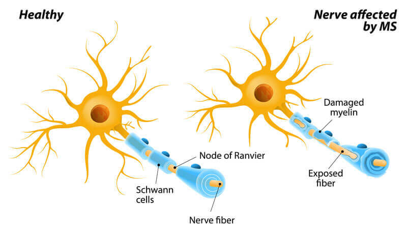
Multiple sclerosis is an autoimmune disease that causes your body to produce antibodies that mistakenly attack the myelin sheath protecting your nerve tissue. This chronic central nervous system disorder damages the nerves and causes the gradual loss of muscle control, strength, and vision.
Multiple sclerosis affects people differently. Some have only mild symptoms, while others are severely debilitated by the disease. Symptoms of multiple sclerosis vary widely and can include the following:
If you are experiencing any of these symptoms, it is important to see your doctor immediately. To determine if you have multiple sclerosis you will be given a neurological examination and, possibly, an MRI scan and other tests to diagnose the cause of your symptoms.
Should your doctor confirm that you have multiple sclerosis there are a number of treatment options. There are several medications that can help, as can physical therapy, occupational therapy, and other treatments.
Although there is no cure for multiple sclerosis the major causes of vision problems associated with the disease are all treatable and often resolve on their own. Three common visual problems associated with MS are:
Steroid medications are commonly prescribed for all three conditions. Patching, prism eyeglasses, and perhaps surgery are also effective in treating double vision. Nystagmus may respond to some medications other than steroids, as well.
Myasthenia gravis is a disorder characterized by weakness of the muscles that are under your voluntary control. Myasthenia Gravis is characterized by a communication breakdown between your nerves and muscles. It can be caused by an autoimmune condition that has damaged the receptors on your muscles. Patients with myasthenia gravis produce antibodies that adhere to the muscle receptors and prevent nerve impulses from getting to the muscle. This causes the muscle to become weakened.
Myasthenia most often affects the muscles of the face, eyes, arms, and legs, as well as the muscles used for chewing, swallowing, and talking. The muscles that control breathing and swallowing can sometimes be involved as well. Some of the signs of myasthenia gravis include:
The symptoms of myasthenia can worsened by fatigue, stress, illness, and certain medications. Check with your doctor before taking any new prescription or over-the-counter medications.
Your ophthalmologist can test for myasthenia using a number of methods, including:
There is no known cure for myasthenia, but if you seek treatment early when you first experience symptoms, you can manage the condition successfully. Your ophthalmologist has a number of treatment options to manage your condition, including medication and surgery. You can also receive physical therapy and learn specific coping skills to help improve your daily life. Early detection and treatment of myasthenia is crucial to managing the condition and preventing serious problems with breathing or swallowing (which require emergency care).
Optic neuritis is a condition characterized by inflammation of the optic nerve. This nerve is the pathway that carries impulses from the retina in the back of the eye to the brain and enables the brain to interpret the impulses as images. If the nerves are damaged, vision is greatly affected.
This condition may affect one or both eyes, and symptoms may appear slowly or over a few days. Some of these symptoms include blurred or dim vision, abnormal color vision, or pain in the back of the eye socket or when moving the eyes. These symptoms may get worse with heat or exhaustion. If you are experiencing any of these symptoms, see your ophthalmologist for an eye examination. If optic neuritis goes untreated, symptoms will get worse.
The causes of optic neuritis are known to be associated with various diseases such as mumps, influenza, measles, multiple sclerosis, or vascular occlusions. However, in many cases, optic neuritis occurs with no known cause.
Steroid drugs are sometimes used to treat optic neuritis. In most patients, vision will significantly improve or return to normal without treatment.
Orbital inflammatory pseudotumor is characterized by inflammation within the orbit, or eye socket, that mimics symptoms similar to a tumor in the same site. The cause is unknown.
Orbital inflammatory pseudotumor usually occurs in only one eye. Symptoms may include:
Your ophthalmologist will probably order a scan to confirm the diagnosis In order to rule out other conditions, your ophthalmologist may run other tests and biopsy orbital tissues if necessary.
Orbital inflammatory pseudotumor is usually treated with steroid medications. If further treatment is necessary, radiation therapy is another option. In some cases the symptoms may return so regular monitoring of the condition is necessary.
Pseudotumor cerebri (PTC) or idiopathic intracranial hypertension (IIH) is a condition in which the pressure from the cerebral spinal fluid inside your head is elevated. This can cause problems such as headaches, blurred vision, or loss of vision. The condition is known as pseudotumor cerebri because symptoms can mimic those of a tumor.
The cerebral spinal fluid (CSF) is a clear fluid that bathes the brain and spinal cord. In cases of PTC, the pressure in this fluid is elevated. The pressure causes swelling of the optic nerve (in the back of the eye) which can lead to loss of vision. It can also damage the nerves that control eye movement, resulting in double vision.
The causes of PTC are not certain, but they may include the following:
The most common symptoms of PTC are headache and visual loss. The headache can be located anywhere, but is usually in the back of the head. It may wake you in the middle of the night, and it may worsen with bending or stooping. Other symptoms include:
Your ophthalmologist will give you a complete eye examination. It may be necessary for you to have an MRI scan and spinal tap to assure accurate diagnosis and to rule out other abnormalities of the cerebrospinal fluid.
If your symptoms are mild, no treatment other than weight loss and careful monitoring may be necessary. Often medications (diuretics) can help lower CSF pressure.
If your vision continues to deteriorate after you have begun treatment, surgical procedures may be undertaken in order to protect the optic nerves from any further damage.
A stroke is a life-threatening emergency in which the blood supply to the brain is interrupted or severely reduced, depriving it of oxygen and killing brain cells. Quick treatment could save your life and minimize damage to your brain.
If you are experiencing these symptoms, seek immediate medical attention. Receiving treatment within three hours of suffering a stroke is shown to dramatically improve your chances of a successful recovery.
There is no treatment for patients who have lost vision due to a stroke. However, you may regain some of the peripheral vision lost from a stroke. Your ophthalmologist will give you a thorough eye examination to determine how the stroke has affected your vision. He or she will talk to you about what to expect over time and can help you find resources and training to make the most of your remaining vision.
Traumatic optic neuropathy is the sudden, severe loss of vision following blunt injury to the eye or areas surrounding the eye. The optic nerve can be damaged by the blow itself, or as a result of other damage sustained by the eye. Vision loss can be immediate or may take days, weeks, or even months to develop.
Your ophthalmologist will give you a thorough eye examination, and you will receive a neurological examination as well, especially if you lost consciousness after the injury. An MRI or CT scan will confirm the diagnosis of traumatic optic neuropathy and verify that no other damage has occurred due to the injury.
If you have mild symptoms, you might only need close observation by your ophthalmologist. Some patients show some improvement with no medical intervention. However, many patients need treatment with corticosteroid medication to reduce the inflammation that is causing vision loss.
Discuss the complications of corticosteroid use with your ophthalmologist.
In some cases, corticosteroids do not fully resolve the condition. In these cases, your ophthalmologist may recommend optic nerve decompression surgery. If your ophthalmologist thinks this a valuable treatment option for you, discuss the benefits and risks together before deciding on surgery.
Visual field testing is a critical part of the neuro-ophthalmic exam and is essential for the evaluation of unexplained visual loss. A visual field test measures all areas of your eyesight, including your side, or peripheral, vision. This crucial test helps your ophthalmologist tell whether there are gaps in your vision. It also helps diagnose your condition, as the test can help find certain patterns of vision loss that may rule out certain conditions or help specify the source of your vision loss.
To take this painless test, you sit facing a bowl-shaped instrument called a perimeter. While you stare at the center of the bowl, lights flash. Each time you see a flash, you press a button. A computer records the location of each flash and whether you pressed the button when the light flashed in that location. At the end of the test, a printout shows if there are areas of your field of vision where you did not see the flashes of light. This test shows if you have any areas of vision loss.
Regular perimetry tests are a key way to see how, if at all, your vision is changing over time. It can also be used to see if your treatments are successful at improving your vision or preventing further vision loss.