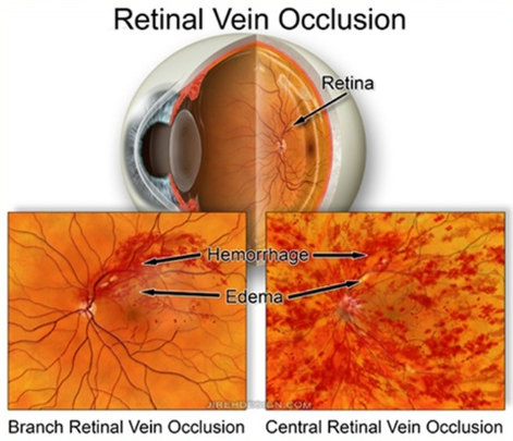
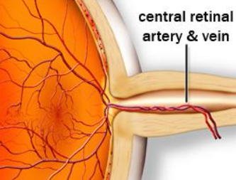
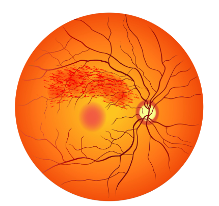
A retinal vein occlusion (RVO) is a blockage in one of the veins that drain blood from the retina, the light-sensitive tissue at the back of the eye responsible for vision. When one of these veins becomes obstructed, blood and fluid can leak into the retina, leading to swelling, bleeding, and vision changes.
There are two main types of retinal vein occlusions:
Retinal vein occlusions are most often caused by changes in the retinal blood vessels, such as thickening or hardening of the vessel walls. Risk factors include:
In some cases, blood clotting disorders may also contribute to the development of an occlusion.
Patients may experience sudden, painless vision loss or blurring in one eye. The severity of visual impairment depends on the location and extent of the blockage. In some cases, only part of the vision is affected. In others, central vision can be significantly reduced.
Diagnosis is made through a comprehensive dilated eye examination. Additional imaging tests, such as optical coherence tomography (OCT) and fluorescein angiography, may be performed to evaluate retinal swelling and blood flow.
While the blocked vein itself cannot be reopened, treatment focuses on managing complications and preserving vision. Common approaches include:
Visual outcomes vary depending on the severity of the occlusion and how promptly treatment begins. With appropriate management, many patients can maintain or recover useful vision. Ongoing follow-up is important to monitor for recurrence or complications such as macular edema or neovascularization.